Single-cell studies are a valuable part of an integrative multiomics research approach to discover new cell types, capture cellular diversity, and understand cellular functions – information that can help resolve disease mechanisms, identify therapeutic targets, and understand epigenomic regulation.
The accuracy and reproducibility of single-cell analysis, however, can be directly impacted by the quality of the starting sample, therefore it is critical to prepare high-quality, single-cell or single-nuclei suspensions from tissue samples. Here, we describe the characteristics of high-quality single-cell and single-nuclei suspensions and discuss factors that may influence sample quality.
What constitutes a high-quality single-cell suspension?
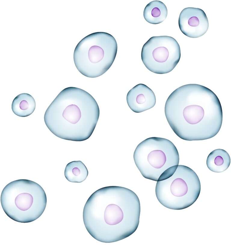
High-quality single-cell suspensions consist of intact, viable, and functional cells that are of the size and shape consistent with their expected morphologies and in the expected proportions for the tissue of origin. Moreover, high-quality suspensions exhibit minimal cell aggregation, low levels of debris, and are at the appropriate concentration for the downstream assay (Figures 1, 2).
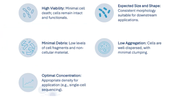
Figure (1) Characteristics of a high-quality single-cell suspension.
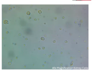
Figure (2) A 40x magnification of kidney cells prepared as a high-quality single-cell suspension using the Singulator™ Platform.
What does a high-quality, single-nuclei suspension look like?
Viable, single-cell suspensions cannot be prepared from OCT-embedded or frozen tissue samples. Instead, researchers can prepare single-nuclei suspensions from these sample types for single-cell analysis. High-quality single-nuclei suspensions contain intact nuclei that maintain a smooth, round shape indicating the retention of nuclear contents and intact heterochromatin structures. The absence of nuclear aggregates and debris in the suspension is also required for successful downstream analysis (Figures 3, 4).
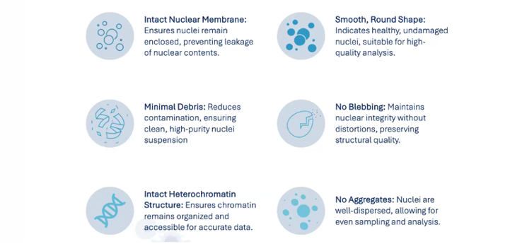
Figure (3) Characteristics of a high-quality single-nuclei suspension.
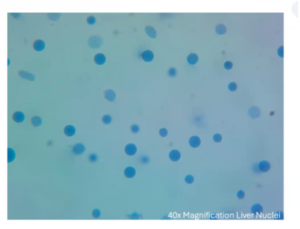
(4) A 40x magnification of a high-quality liver nuclei suspension prepared using the Singulator™ Platform.
What about FFPE samples?
Researchers increasingly recognize the value of FFPE archives and are using them for single-cell genomics assays. One key advantage of working with FFPE preserved tissues is the ability to store rare clinical samples long-term in a safe and accessible format. Many researchers think that unlocking clinical archives for single-cell analysis will enable discoveries that were previously unobtainable using fresh or frozen samples.
Now, researchers can isolate either cells or nuclei from FFPE samples and use these suspensions in assays designed for fixed tissues. While these suspensions don’t need to be completely free of debris (since downstream steps help clear debris before cellular barcoding), they must still consist of singulated cells (Figures 5, 6) or nuclei. Finally, assays compatible with FFPE preserved tissue rely on a probe-based approach, so scientists must use probes that have been designed for use with their species of interest.
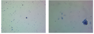
Figure (5) Mouse pancreatic ductal adenocarcinoma FFPE sample, post-cell isolation, 10x magnification, and (6) 40x magnification.
High-quality samples: there’s more than meets the eye
Despite meeting the visual criteria outlined above, single-cell and single-nuclei suspensions may still fail to deliver reliable and reproducible data. Multiple factors can influence gene expression and chromatin accessibility, so it is important to standardize sample preparation to minimize variability. For example, inconsistencies in sample collection, preservation, and isolation methods can influence yields, viability, membrane integrity, chromatin structure, and gene expression. The choice of dissociation method should be considered to ensure compatibility with your research goals. Manual and semi-automated sample dissociation methods, for instance, can be difficult to standardize, introducing technical variation that can make it difficult to recover fragile or rare cell types or compare data across studies.
The value of automated sample preparation
By following best practices for sample collection and storage, along with automated preparation methods, researchers can achieve consistent results regardless of who performs the isolation. The Singulator™ Platform delivers reliable, high-quality data by isolating cell populations with minimal bias, which is crucial for single-cell genomics applications. The Singulator Platform streamlines workflows by reducing manual steps, saving time, and minimizing errors.
Designed for versatility, the Singulator handles very low input masses, down to 2 mg, and works with FFPE samples (Figure 7), making it the most flexible solution available. Additionally, the cell and nuclei suspensions prepared using the Singulator Platform are compatible with a wide range of single-cell genomics applications, including gene expression assays, chromatin accessibility assays, multiome assays, whole genome amplification (WGA), single nucleotide variant (SNV) analysis, and copy number variation (CNV) detection.
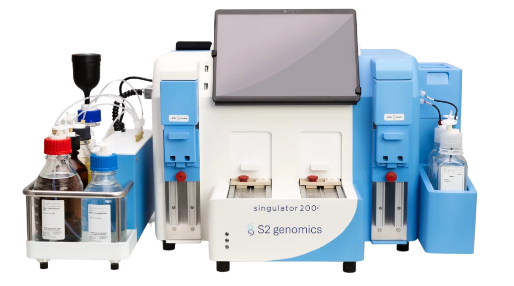
Figure 7. The Singulator™ 200+ System automates FFPE sample processing.
Interested in learning more?
Watch our webinar, “Automating Tissue Dissociation for Single-Cell Analysis,” for more information on how to get started preparing high-quality single-cell and single-nuclei suspensions with the Singulator Platform, or read our brochure.


